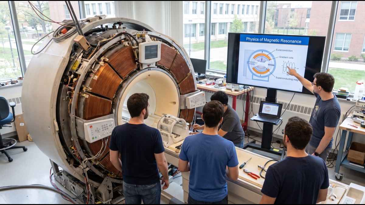
An MRI machine can produce a remarkably detailed picture of the soft tissue inside a human body without a single X-ray and without any ionizing radiation at all. At its physical core, the technology relies on nothing more exotic than a very large magnet, carefully timed radio waves, and the simple chemical fact that the human body is, by mass, mostly water.
Understanding how those three ordinary ingredients combine to build a three-dimensional anatomical image explains not only why MRI scanners look and sound the way they do, but also why certain patients cannot safely enter one, why scans take so long, and why the machine is genuinely one of the more unusual pieces of everyday medical technology once its underlying physics is unpacked.
Why an MRI Needs No Radiation At All
Magnetic resonance imaging works on an entirely different physical principle from an X-ray or a CT scan, both of which build an image by passing ionizing radiation through the body and measuring how much of it is absorbed or blocked by different tissues along the way.
MRI instead exploits a property of the hydrogen atoms already present in enormous numbers throughout the body's water and fat, using strong magnetism and radio-frequency energy to make those atoms emit a detectable signal, a process that involves no radiation exposure whatsoever and is generally considered safe to repeat as often as clinically necessary.
This absence of ionizing radiation is precisely why MRI is often the preferred imaging method for children, for repeated monitoring of a chronic condition over years, and for imaging pregnant patients when a scan is genuinely necessary, situations where minimizing radiation exposure carries particular clinical importance.
Why the Human Body Is Perfect Raw Material
The hydrogen nucleus, a single proton, behaves like a tiny spinning magnet, and the human body happens to be an unusually rich source of these protons because water alone makes up roughly two-thirds of total body mass, with additional hydrogen present throughout fat and other tissue.
In their normal resting state, these countless proton magnets point in essentially random directions throughout the body, and their combined magnetic effect cancels out almost entirely, producing no detectable signal for an external scanner to pick up under ordinary conditions.
Placing the body inside a sufficiently strong external magnetic field changes this picture completely, because a majority of these tiny proton magnets begin aligning either with or against that external field, creating for the first time a measurable net magnetization that the scanner can subsequently manipulate and read.
What the Main Magnet Actually Does
The large cylindrical tube that most people picture when they think of an MRI machine houses an exceptionally powerful magnet, typically many thousands of times stronger than Earth's own natural magnetic field, generating the uniform magnetic environment needed to align the body's hydrogen protons in the first place.
This main magnetic field has to be extremely uniform across the entire area being imaged, since even small inconsistencies would distort the resulting image, which is why the magnet itself represents one of the most expensive and precisely engineered components of the entire machine.
Field strength is measured in a unit called the tesla, and clinical scanners commonly operate between roughly 1.5 and 3 tesla, with even stronger research-grade magnets existing for specialized applications that demand finer image detail than routine clinical scanning requires.
Why the Magnet Has to Be Superconducting
Generating a magnetic field that powerful using an ordinary electromagnet would require so much continuous electrical current that the coil would generate destructive amounts of heat and consume an impractical amount of power simply to stay switched on hour after hour.
Clinical MRI magnets solve this by using superconducting wire cooled to an extremely low temperature using liquid helium, at which point the wire loses essentially all electrical resistance, allowing an enormous current to circulate through the coil indefinitely without generating heat or requiring continuous external power once it has been established.
This is why an MRI scanner is technically never fully switched off between patients, and also why the machine requires periodic replenishment of liquid helium and specialized handling, since a sudden loss of superconductivity, called a quench, causes the stored energy to release rapidly and can be a genuinely significant engineering event.
What Happens When the Radio Pulse Hits
With the body's protons now aligned by the main magnetic field, the scanner briefly transmits a precisely tuned pulse of radio-frequency energy at exactly the resonant frequency those aligned protons will absorb, which knocks a portion of them out of their aligned resting state.
The specific frequency required to achieve this effect depends directly on the strength of the local magnetic field, which is the essential physical link that allows the machine to later determine where in the body a given signal originated, since the field strength is deliberately varied across different locations.
Once the radio pulse switches off, the disturbed protons begin realigning themselves back with the main magnetic field, and as they do so they release the absorbed energy back out as a faint radio signal of their own, which is the actual signal the scanner's receiver coils are built to detect.
How Relaxation Time Creates Image Contrast
Different types of body tissue return to their aligned resting state at measurably different speeds after the radio pulse switches off, a property described using two distinct relaxation time measurements that radiologists refer to as T1 and T2, each sensitive to slightly different physical characteristics of the tissue.
Fat, water, muscle, and abnormal tissue such as a tumor or areas of inflammation each display characteristically different T1 and T2 relaxation behavior, and it is precisely this difference in how quickly various tissues release their absorbed energy that the scanner translates into the varying brightness levels seen in the final image.
Radiologists deliberately choose between T1-weighted and T2-weighted scanning sequences, along with several other specialized sequence types, depending on which tissue characteristics are most diagnostically relevant to the specific clinical question being investigated, which is why a single MRI examination frequently includes several different scan sequences of the same body region.
Why Gradient Coils Make the Loud Banging Noise
A uniform magnetic field alone cannot tell the scanner where within the body a particular signal originated, so additional coils called gradient coils are used to deliberately make the magnetic field strength vary slightly and precisely across different locations within the scanner bore.
Because the resonant frequency protons respond to depends directly on local field strength, this deliberate variation lets the scanner mathematically determine the exact three-dimensional location each portion of the received signal came from, which is the essential trick that allows MRI to build a genuine spatial image rather than a single undifferentiated signal.
The loud banging and clicking noise closely associated with an MRI scan comes directly from these gradient coils physically vibrating as very large electrical currents are switched on and off extremely rapidly throughout the scan, which is why patients are routinely given hearing protection before entering the scanner.
How the Machine Turns Signals Into a Picture
The raw radio signals collected by the scanner's receiver coils do not resemble a recognizable image at all when first captured; they exist instead as a complex mathematical dataset commonly referred to as k-space, representing spatial frequency information rather than direct pixel brightness values.
A mathematical technique called a Fourier transform converts this k-space data into the recognizable cross-sectional image radiologists actually interpret, a computationally intensive step that modern scanner hardware performs essentially in real time as data continues to be collected throughout the scan.
Each individual image represents a thin anatomical slice through the body, and a complete examination typically collects many such slices across the region of interest, which the software can subsequently combine into a three-dimensional reconstruction that can be viewed from angles beyond the original scanning planes.
What Contrast Dye Actually Changes
Some MRI examinations use an injected contrast agent, most commonly one containing the element gadolinium, to improve the visibility of specific structures such as blood vessels, areas of inflammation, or certain tumors that might otherwise be difficult to distinguish clearly from surrounding normal tissue.
Gadolinium works by altering the local magnetic environment immediately surrounding it, which changes how quickly nearby hydrogen protons relax after the radio pulse, effectively brightening or darkening specific tissue on the resulting image depending on how much contrast agent has accumulated in that particular location.
Because gadolinium-based agents carry their own specific safety considerations, particularly for patients with significantly reduced kidney function, radiologists weigh the diagnostic benefit of improved contrast against these considerations on a case-by-case basis rather than using contrast agents in every examination as a matter of routine.
Why MRI Excels at Soft Tissue Over Bone
MRI is generally considered the superior imaging method for soft tissue structures such as the brain, spinal cord, muscles, ligaments, and internal organs, precisely because these tissues contain abundant hydrogen-rich water and fat that generate a strong, richly differentiated signal.
Dense bone, by contrast, contains comparatively little mobile water and therefore generates a much weaker MRI signal, which is why X-rays and CT scans, which rely on a completely different physical mechanism better suited to dense material, generally remain the preferred imaging method for evaluating fractures and bone structure directly.
This complementary relationship is why a single patient with a complex musculoskeletal injury might reasonably receive both an X-ray to evaluate bone and an MRI to evaluate the surrounding soft tissue, since neither technology alone provides a complete picture of every relevant structure.
Why Certain Implants Make MRI Dangerous
The enormous magnetic field at the heart of an MRI scanner is strong enough to forcefully move, heat, or dislodge any object made from ferromagnetic metal that enters the scanning room, which is why metal objects such as tools, oxygen tanks, and even furniture must be rigorously excluded from the area.
Certain medical implants pose a similarly serious risk, since some older pacemakers, specific aneurysm clips, and various other metallic devices can be moved, heated, or electrically disrupted by the scanner's magnetic and radio-frequency fields, which is why patients are always carefully screened for implants before any scan proceeds.
Many modern implanted devices are now specifically manufactured and certified as MRI-conditional or MRI-safe under defined scanning conditions, but verifying the exact make, model, and safety status of any implant remains an essential step that radiology staff complete before allowing a patient into the scanning room.
Why Some Patients Struggle With the Scanner Itself
The scanning tube itself is narrow and enclosed, and a standard examination can require a patient to remain still inside it for anywhere from roughly fifteen minutes to well over an hour depending on how many sequences the specific examination requires.
This combination of enclosed space, prolonged stillness, and loud repetitive noise causes genuine claustrophobia or significant anxiety for a meaningful proportion of patients, which has driven the development of wider-bore scanner designs and, for some facilities, open MRI configurations specifically intended to reduce this discomfort.
Sedation is occasionally used for patients who cannot otherwise tolerate the examination, particularly young children and severely claustrophobic adults, though this adds its own logistical and medical considerations that facilities weigh carefully against the diagnostic necessity of the scan.
What Functional MRI Adds to the Picture
A specialized variant called functional MRI extends the same underlying physics to detect not just anatomical structure but ongoing brain activity, by measuring subtle changes in blood oxygenation that occur in brain regions working harder at a given moment.
This works because oxygenated and deoxygenated blood have measurably different magnetic properties, meaning a brain region receiving increased blood flow during active mental tasks produces a detectably different signal from the same region at rest, without requiring any additional contrast agent or radiation.
Functional MRI has become a significant tool in neuroscience research and in specific clinical applications such as presurgical mapping of critical brain regions before a tumor operation, though interpreting its results correctly requires considerable statistical and methodological care that remains an active area of ongoing research refinement.
Why a Scan Takes So Long to Complete
Building a single detailed cross-sectional image requires the scanner to collect and process a substantial amount of signal data across a repeated sequence of radio pulses, and a complete examination typically requires many separate sequences to capture different tissue contrasts and anatomical planes.
This is fundamentally different from a single instantaneous X-ray exposure, and is the core reason MRI examinations routinely take considerably longer than other forms of medical imaging, a genuine tradeoff for the substantially greater soft-tissue detail and complete absence of radiation exposure the technology provides.
Newer scanning sequences and more powerful computing hardware have meaningfully reduced typical scan times over recent years, and ongoing research continues to push toward faster acquisition without sacrificing the image quality that makes MRI diagnostically valuable in the first place.
What looks from the outside like a simple if imposing cylindrical machine is, underneath its cover, an elegant application of nuclear magnetic resonance physics repurposed for medicine: aligning the body's own abundant hydrogen protons with a powerful magnetic field, briefly disturbing that alignment with a precisely tuned radio pulse, and then carefully listening to how differently each type of tissue returns to its resting state.
That entire chain, from magnet to radio pulse to relaxation signal to reconstructed image, is why an MRI scanner can see deep inside the body with extraordinary soft-tissue detail using nothing that resembles radiation at all, and why so many of its practical quirks, from the noise to the screening questions to the length of the appointment, trace directly back to that same underlying physics.
Sources
- Wikipedia — overview of magnetic resonance imaging technology and physics
- National Institute of Biomedical Imaging and Bioengineering (NIH) — patient and clinical information on MRI
- RadiologyInfo.org (RSNA/ACR) — public radiology education resource on MRI procedures and safety
- U.S. Food and Drug Administration — MRI safety guidance and contrast agent regulation
- World Health Organization — medical imaging and diagnostic technology resources
FAQ
Does an MRI scan use radiation?
No. MRI uses a strong magnetic field and radio waves rather than ionizing radiation, which is a fundamental difference from X-rays and CT scans.
Why is an MRI machine so loud?
The banging noise comes from gradient coils physically vibrating as large electrical currents switch rapidly on and off to encode spatial position in the image.
Why can't people with certain implants have an MRI?
The magnetic field is strong enough to move or heat ferromagnetic metal objects and can disrupt electronic devices like some pacemakers, so implants must be verified as MRI-safe beforehand.
What is contrast dye used for in an MRI?
A gadolinium-based contrast agent alters how nearby tissue responds to the magnetic field, making certain structures like blood vessels or tumors stand out more clearly.
Why does an MRI scan take so long?
Building a detailed three-dimensional image requires collecting many individual signal readings across repeated pulse sequences, which is inherently slower than a single instantaneous X-ray exposure.
About the Author
We reference Wikipedia, the National Institute of Biomedical Imaging and Bioengineering, RadiologyInfo.org, the U.S. Food and Drug Administration, and the World Health Organization to explain the background and current understanding of this topic.
Loved This Article?
Share it on WhatsApp → Share it on WhatsApp
Get more guides in your inbox — Subscribe to our newsletter for weekly surprising stories from Egypt, Saudi Arabia, Dubai, and beyond.